Scientists Capture Crispr’s Gene-Cutting in Action
Scientists from the University of California, Berkeley have detailed how Cas9 changes the structure of its target DNA to position each strand for cleavage. In their paper published in the journal Science on Jan 14 2016, the team describes their study of the way three-stranded structures known as R-loops are formed during the technique and how they used a familiar technique to capture 3D images of the process and what they learned as a result.
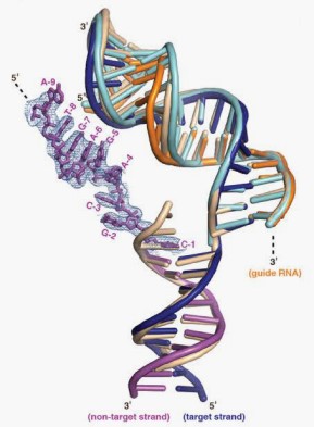
The structures help explain how Cas9 holds unwound double-stranded DNA, which enables the formation of an RNA-DNA hybrid, without ATP-dependent helicase activity. "DNA binding at a sequence complementary to the 20-nt guide RNA segment in the Cas9–RNA complex induces protein structural rearrangements that accommodate both the RNA–DNA helix and the displaced non-target DNA strand," the authors wrote. "Those protein–nucleic acid interactions in turn direct the non-target DNA strand into the RuvC domain active site, favoring local conformational changes that position the HNH domain active site near the scissile phosphate of the target DNA strand." The protein induces a 30 degree bend in the helix that stabilizes the R-loop and allows the nuclease domains to cleave the double-stranded DNA.
This website stores cookies on your computer. These cookies are used to collect information about how you interact with our website and allow us to remember you. To find out more about the cookies we use, see our Privacy Policy .